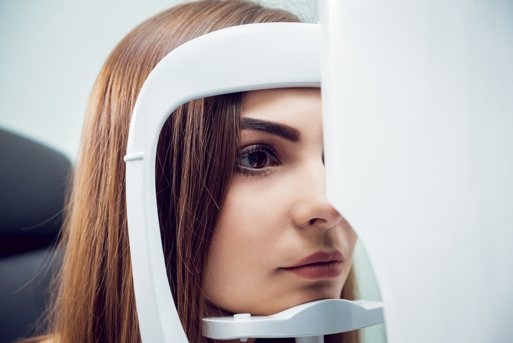
Common Imaging Modalities in Ophthalmology
Improvements in image processing and computer hardware have led to the advancement of existing imaging modalities and the creation of novel imaging techniques that have transformed ophthalmic patient care.
The creation of the Scanning Laser Ophthalmoscope (SLO) and Optical Coherence Tomography (OCT) has revolutionized ophthalmology, and clinical trials have supported their robust efficacy and diagnostic utility in many diseases of the eye. New imaging modalities such as adaptive optics (AO) have improved the histologic study of single cells in the retina and anterior segment. The emergence of telehealth has led to the creation of handheld OCT devices and imaging through smartphones. These new imaging modalities will pave the way for developing novel therapeutic approaches, requiring ophthalmology CROs to initiate clinical trials for these therapeutics.
In this article, we will discuss the common imaging modalities in ophthalmology, current novel imaging approaches, and explore the developing research behind telehealth imaging modalities that will likely revolutionize the ophthalmology industry.
Original Ophthalmic Imaging Modalities
Less than 40 years ago, eye imaging was limited to external photography, slit-lamp photographs, fluorescein angiography, fundus and retinal photographs, and specular microscopy. These techniques have come a long way since their original invention and are still used in regular practice today. Technological advances have simplified and streamlined their use for practicing ophthalmologists.
External Eye Photography
Although this may seem obvious, external eye photography provides invaluable information on the external appearance of the eyes and surrounding lid and facial structures. Conventional macro photography using digital single-lens reflex cameras is used to demonstrate facial nerve anomalies, record eye or surrounding tissue lesions, and document surgical alignment of the eyes or eyelids. Motility photographs may also be taken to record positions of the eyes together and in various gazes to evaluate for the misalignment of eyes due to issues with extraocular muscles.
Slit-Lamp Photography
Slit-lamp photography is used for imaging the anterior segment of the eye. The photo slit-lamp is an adaptation of the slit-lamp biomicroscopy used in ophthalmology for lighting and examining the eye. Photo slit-lamps use beam splitters to direct the light to a camera-mounted instrument. These photos allow for examination of the anterior segment of the eye, including the cornea, lens, and iris. It is often used to diagnose damage, inflammation, blockage, and vision loss.
Retinal or Fundus Photography
Retinal or fundus photography is a fast and straightforward procedure. Fundus photography requires pupil dilation with short-acting mydriatic eye drops. Photographs of the posterior segment of the eye will be captured and include images of the retina, optic nerve head, macula, retinal blood vessels, choroid, and vitreous. These images are used to screen and diagnose various treatable eye conditions causing blindness, such as diabetic retinopathy, age-related macular degeneration, glaucoma, retinopathy of prematurity, and other conditions impacting the retina.
During the last ten years, non-dilated cameras have been developed and are particularly useful in retinal screening programs when many people require imaging without pupil dilation. Non-mydriatic cameras are vulnerable to opacity effects from diseases like cataracts, which may impact imaging quality. Mydriatic cameras are currently the imaging modality of choice, especially if the peripheral retina requires imaging.
Fluorescein Angiography
Ophthalmologists have relied on fluorescein angiography (FA) for over four decades to understand, diagnose, and treat retinal disorders. FA has also been used to visualize neovascular vessels at the iris, anterior chamber, or cornea level.
Typically, this invasive diagnostic imaging procedure utilizes a retinal camera or a scanning laser ophthalmoscope to capture retinal and choroidal circulation photographs following intravenous injection of fluorescein sodium. Another fluorescent dye, indocyanine green, has also been used for retinal and choroidal angiography. Images are taken every few seconds as the dye travels through the vessels. Photographs highlight abnormal blood vessels, fluid leaks in the retina, and evaluate the general strength of circulation.
FA aids in diagnosing various ocular pathologies, including retinal pigment epithelium rips, central serous chorioretinopathy, malignant melanoma, choroidal metastasis, and central retinal vein occlusion. This technique is advantageous in the management of diabetic retinopathy and macular degeneration. Fluorescein angiography of the iris or other anterior structures may also be of diagnostic value.
Specular Microscopy
Specular microscopy is a variation of slit-lamp photography and a non-invasive imaging modality that uses a special camera to photograph an individual cell layer of the cornea. The high-magnification specular micrograph images are crucial in assessing the health of the cells. Specular microscopy images help to assess preoperative cell health before high-risk surgeries, evaluate donor cornea before transplantation, compare various surgical techniques, and identify the impact of lasers during refractive surgery.
Traditionally, these instruments required contacting the cornea, but the newest use of non-contact microscopy with semi-automated digital analysis of the cell counts and morphology makes diagnosis much more straightforward.
Ultrasound of the Eye
Ocular ultrasound examination is an important imaging modality for the ophthalmologist. Ultrasound imaging has many potential benefits in evaluating patients with acute vision loss, ocular trauma, headache, or increased intracranial pressure. This examination is painless, fast, and can provide very detailed information for the provider.
The ultrasound can perform specific examinations such as the A-scan or B-scan. A-scan is used to determine eye length, usually for calculating intraocular lens power inserted during cataract surgery. B-scan is used for diagnosing lesions of the posterior segment of the eye, including cataracts, retinal detachment, vitreous degeneration, retinoblastoma, ocular trauma, and choroidal melanoma.
New Imaging Modalities
New imaging modalities in the field of ophthalmology have revolutionized the industry and how ophthalmologists diagnose disease, document the progression of pathological conditions, and monitor treatment response. Common modalities include optical coherence tomography (OCT) and scanning laser ophthalmoscope (SLO). The creation of fluorescence lifetime imaging ophthalmoscopy (FLIO) and adaptive optics (AO) will be influential in the coming years.
Optical Coherence Tomography
Optical coherence tomography (OCT) is a recent imaging procedure helpful in diagnosing several retinal disorders traditionally imaged with fundus photography or FA. This imaging modality uses a computer to evaluate the patterns of light reflected inside the eye. This creates an image similar to the cross-section of retinal tissue that was previously examined under a microscope. This image was impossible to obtain in the living eye until recently.
OCT is used to capture images of the macula, retinal nerve fiber layer, and optic nerve for measurement and subsequent evaluation in detecting retinal diseases and glaucoma. It is beneficial in detecting retinal conditions associated with fluid leakage, including macular holes, cystoid macular edema, subretinal fluid, and retinal pigment epithelial detachments. One of its greatest values is its ability to monitor changes in retinal thickness due to macular edema from diabetic retinopathy or other causes.
The latest iterations of the OCT include OCT angiography, visible light OCT, Full-Field OCT, anterior segment OCT, whole eye OCT, and intraoperative OCT. These iterations allow for a more comprehensive evaluation of patients and provide additional detail on ophthalmologic disease processes.
Scanning Laser Ophthalmoscope
The scanning laser ophthalmoscope is an imaging technique that utilizes a collimated laser beam for viewing the eye. The SLO produces tomographic imaging of ocular structures such as the retina, choroid, optic nerve head, and retinal nerve fiber layer. The technology of the SLO is used in various newer imaging modalities, including scanning laser polarimetry, AO, ultra-widefield imaging, and multicolor imaging that have transformed the diagnosis and treatment of retinal diseases. SLO also captures optic nerve head measurements in patients with glaucoma.
Fluorescence Lifetime Imaging Ophthalmoscopy
Fluorescence lifetime imaging ophthalmoscopy (FLIO) is an emerging imaging modality for in vivo measurement of lifetimes of endogenous retinal fluorophores. The new technique of FLIO measures the decay lifetime of retinal fluorophores, which are unique to molecules and more sensitive to weak fluorophores, and that might be hidden when using fundus autofluorescence. FLIO can reveal structural, metabolic, and biochemical changes in the retina.
FLIO patterns have been identified for a variety of retinal diseases and can be valuable in early detection and detailed monitoring, such as age-related macular degeneration, diabetic retinopathy, vascular occlusive diseases, CSR, choroideremia, macular holes, and is believed to be a promising diagnostic method.
Adaptive Optics
Adaptive optics (AO) is a groundbreaking technology in the biotech field. This retina imaging technique implements wavefront technology that was first utilized in astronomy and defense systems to correct for ocular anatomical and physiological anomalies that glasses, contact lenses, or refractive surgery can’t ameliorate. AO allows in vivo examination of the photoreceptors and retinal vasculature, which may help providers better understand retinal pathologies.
The current imaging modalities for retinal diseases can only detect these pathologies after visible fundus changes occur. AO may aid in recognizing retinal disease before this significant damage occurs, which can help support the progress of ophthalmic disease drug development. AO is presently limited to research because specific barriers such as cost and availability must be overcome before implementation in daily practice.
Telehealth Imaging
Telemedicine has revolutionized the healthcare industry since the COVID pandemic. In response to its growing demand, ophthalmologists have started to employ telemedicine imaging techniques and technologies to better facilitate patient care in a remote setting.
The most common way this is done is by obtaining eye imaging with a smartphone, known as smartphone imaging (SI). Lord et al. (2010), demonstrated that an iPhone could be used to capture external photos of the orbit and surrounding structures and indirectly image the anterior segment and the fundus of the eye when used with a fitted slit lamp. Now, novel external attachments and phone applications have been introduced to improve SI’s image quality and utility. SI has also been utilized in screening common ophthalmologic diseases, such as age-related macular degeneration, glaucoma, and diabetic retinopathy. As this technology advances, implementation in everyday ophthalmic practice will increase in the coming years.
Partner with Vial
The continued advances in the field of ophthalmology imaging, highlight the potential for novel therapeutics that may utilize these imaging modalities to evaluate their efficacy, safety, and tolerability. Additionally, with the guidance of our data science experts at Vial Ophthalmology CRO, you can build an efficient, cost-effective, patient-friendly ophthalmology clinical trial. Contact a team member today to discover how we can help maximize impactful data collection and propel your role in the biotechnology field toward success.


