
In medical school, I was taught that taking a thorough history was enough to lead to a diagnosis in many cases. I was also taught that I could achieve a similar result through a thorough physical examination even if a patient provided no history. While both are true, a good practitioner relies on both.
What Is a Thorough Examination for Dry Eye?
Without trying to make everyone a doctor, it is useful to understand what a thorough physical examination for dry eyes includes. Discussion of what constitutes a detailed history is better directed to eye care providers than to patients.
Many systemic diseases are associated with dry eyes, so examination of a patient’s hands is essential, creating opportunities to spot arthritic changes, such as are seen in Rheumatoid arthritis, that could relate to dry eye. Similarly, examining the skin is vital, allowing eye care providers to seek out signs of conditions that may connect to dry eye, such as rosacea, which often has characteristic facial redness, psoriasis, or allergic diseases.
Patients are people, not just eyes!
Eye doctors are taught that it is important to look at your patient in good lighting first and not to enter a darkened room and proceed directly to the eye examination using the slit lamp microscope that most think of as the eye examination. Patients are people, not just eyes! By beginning the exam with good lighting, it’s possible to evaluate the whole person.
Verifying proper closure of the eyelids comes before examination of the lid anatomy, margin, and oil glands. The quality of the oils produced by the lid meibomian glands must be evaluated, usually by pressing over the glands with a finger or cotton swab.
Eyelashes may show crusting or the presence of germs, even parasites. The conjunctiva may reveal inflammation or even scarring that indicate a cause of symptoms. The cornea (the clear window through which we see) is central to an examination for dry eyes and may show subtle surface changes that indicate a tear film abnormality.
Certain “vital dyes” can be dropped onto the eye surface to reveal “staining” or evidence of improper health of the surface of the eye. In-office tests of tears can indicate abnormal osmolarity (“saltiness” is the simplest way to explain this complex term) or abnormal inflammation that are common in dry eyes.
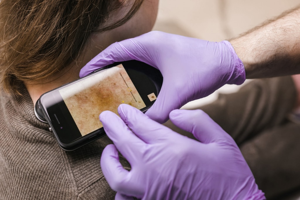
Special photography or imaging of the eyelid oil glands can reveal abnormalities in oil production – very common in patients with dry eye. Such imaging (meibography) has dramatically improved the ability of eye care providers to properly diagnose eyelid-caused dry eye problems. Tear production can be measured using the “Schirmer test,” which applies a small piece of absorbent paper to the surface for five minutes, directly measuring tear production.
Diagnosis comes before treatment
Since there are different causes of dry eyes, it is necessary to thoroughly identify the specific causes in each patient before making treatment recommendations. After all, what works wonderfully for some may not work at all for others, particularly if you’re targeting the wrong core issue.
Follow Vial on LinkedIn for More Information
As part of the medical research community, Vial aims to make critical information accessible. Follow us on LinkedIn to stay up-to-date on everything in the clinical trials industry.
![A companion diagnostic (CDx) is “a medical device, often an in vitro diagnostic (IVD), which provides information that is essential for the safe and effective use of a corresponding drug or biological product”[1]. IVDs refer to lab tests on samples of bodily fluids, e.g., blood, urine, and saliva, to diagnose, monitor, and manage diseases and conditions.](https://vial.com/wp-content/uploads/2023/10/shutterstock_1826989610.jpg)
