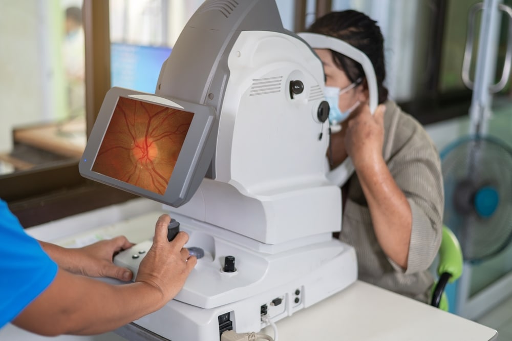
Advancements in retinal imaging techniques have revolutionized the field of ophthalmology, enabling more accurate diagnoses and enhanced monitoring capabilities for various ocular diseases. This article explores the top five innovative techniques for imaging the retina, highlighting their unique features and clinical applications. From Optical Coherence Tomography Angiography (OCTA) to Molecular Imaging with the DARC approach, these cutting-edge technologies provide valuable insights into the retinal structure, function, and molecular changes. By harnessing the power of these techniques, ophthalmologists can improve patient outcomes and develop novel therapeutic interventions for retinal diseases.
Trending Top Retinal Imaging Techniques and Innovations
These innovative imaging modalities have revolutionized the way eye clinics diagnose and monitor ocular diseases. Let’s explore the top five trending retinal imaging techniques and innovations that are shaping the future of ophthalmic care.
1. Optical Coherence Tomography Angiography (OCTA)
Optical coherence tomography (OCT), which has been in use for 25 years, still continues to boast large-scale adoption by eye clinics around the world, despite challenges related to cost and device training.
Impressively, OCT systems have expanded to become even more essential as a diagnostic tool. In recent years, many advances have been applied to the OCT platform with novel improvements in the image quality, and speed of acquisition, setting the stage for more functional extensions of OCT, such as OCT angiography (OCTA).
OCTA is characterized by the latest non-invasive methodologies to enter this space which showcases the ability to take a volumetric angiographic image in mere seconds. Impressively, it works by employing motion contrast imaging to high-resolution volumetric blood flow information generating, which allows for lightning-fast image capture. By comparing the inverse correlation differences between a sequence of OCT scans taken at the same cross-section, it can map the volumetric blood flow. Useful in many diverse diagnostic applications, OCTA imaging techniques are used in evaluating the presence of ophthalmologic diseases such as glaucoma, diabetic retinopathy, artery and vein occlusions, and age-related macular degeneration (AMD).
OCTA is often referenced in comparison to its more invasive counterpart, Fluorescein angiography (FA) and indocyanine green angiography (ICGA). ICGA has always been the gold standard for diagnostic use, however, OCTA demonstrates significant advantages such as:
- 3D OCT data allows for a more complete vantage point of structural information about the retina.
- In contrast, FA is limited to capturing only a 2D view.
- Clinical trials find that OCT works well in tandem with other methodologies listed below.
- As a technology, OCT can be leveraged effectively to produce the most valuable information leading to more favorable patient outcomes, especially when used in conjunctive applications.
2. Photoacoustic Imaging (PAI)
Photoacoustic imaging (PAI) is also referred to as optoacoustic imaging and it is a novel, hybrid, non-invasive imaging technology that captures high-definition images. Dually focused, PAI combines spectroscopic-based specificity and light-induced sound wave measurements produced by optical excitation.
The PAI hybrid process allows for in-depth ocular characterizations by providing high contrast and definition-rich imagery and is a highly-effective technique for identifying abnormalities in the vasculature of the retina. PAI is often used in tandem with other imaging modalities, such as scanning laser ophthalmoscopy (SLO), fluorescence microscopy (FM), and optical coherence tomography (OCT) to gain a deeper perspective of any ocular disease presence and progression.
3. Scanning Laser Ophthalmoscopy (SLO) – Adaptive Optics
Modern confocal SLO utilizes a scanning laser ophthalmoscope to deliver a visible light or near-infrared radiation laser beam across the retina and collect light from each retinal spot as it is illuminated, producing high contrast, reflectance images using the small diameter, centered apertures (confocal apertures). This technology, combined with adaptive optics, has the ability to yield the type of qualitative clinical trial data capable of generating stronger diagnostic criteria and biomarkers for advanced retinal disease.
By providing cellular and subcellular details without the need for histology, scientists can use AO in clinical studies to better comprehend eye changes over time. Also, considered to be a non-invasive approach, AO retinal imaging is safe and easily tolerated by patients. Clinical trials involving AOSLO techniques work to discover cellular-specific approaches to designing novel therapeutic interventions and enhanced monitoring capabilities.
4. Ultra-Widefield Fundus Autofluorescence Imaging (UWF-FAF)
Fundus autofluorescence (FAF) is a technique that maps fluorophores within the ocular structures, which essentially may indicate the presence of disease, whereas ultra-widefield fundus autofluorescence (UWF-FAF) helps to observe the entire retinal periphery in rich detail. Comparatively, predecessor models of fundus imaging only allowed for up to 50% of the retinal periphery view, which compromised the potential for a more complete diagnostic picture or progression analysis.
Fundus Autofluorescence (FAF)
FAF utilizes the natural autofluorescent properties of lipofuscin to detect its presence or buildup, providing insights into the metabolic status and overall well-being of the retinal pigment epithelium (RPE).
When analyzing FAF images, areas of hypoautofluorescence indicate dark regions where no lipofuscin is detected. This could suggest RPE cell death, leading to vision loss or scotomas. Alternatively, hypoautofluorescence may be caused by signal absorption due to factors like blood, pigment, or other blocking artifacts. Common observations associated with hypoautofluorescence include RPE atrophy, new hemorrhages, exudative lesions, laser scarring, dense hyperpigmentation, forms of hard drusen, and vitreous opacities.
On the other hand, hyperautofluorescent areas appear brighter than the normal gray background autofluorescence. These regions indicate an excess accumulation of lipofuscin, potentially reflecting increased metabolic activity of the RPE.
In summary, FAF does not require the injection of a fluorescein dye in order to image the retina but rather utilizes the fluorescent properties of lipofuscin within the RPE to create an image. FAF imaging is an excellent tool as both a diagnostic and monitoring modality for the progression of retinal dystrophies.
The American Academy of Ophthalmology reveals as a standalone, FAF methodology boasts a strong 95% accuracy in its diagnostic capacity for Stargardt disease, retinitis pigmentosa, and Best disease. Additionally, it has clinical application value for a myriad of other advanced eye conditions, including:
- Geographic atrophy, particularly in advanced non-exudative age-related macular degeneration
- Central Serous Chorioretinopathy
- Retinitis pigmentosa and rod-cone dystrophies
- Stargardt disease
- Best disease and vitelliform maculopathies
- Central areolar choroidal dystrophy
- Pattern dystrophies
- Hydroxychloroquine retinopathy and other retinal drug toxicities
- Choroidal nevi and melanomas
- White Dot Syndromes
- Fundus flavimaculatus
Ultra-Widefield Fundus Autofluorescence Imaging (UWF-FAF)
UFW-FAF in clinical research is a focal point for leading CRO teams to help uncover new clinical applications. For example, patients with diabetic retinopathy tend to experience delays in diagnosis because alterations within the retina aren’t easily detected through color imaging. Whereas, UFW-FAF demonstrates high viability toward detecting early molecular changes associated with diabetic macular edema, retina detachment, and other forms of advanced retinopathy. As an adjunctive imaging modality, UFW-FAF is ideal because it facilitates reproducible evidence in evaluating retinal detachment, pre and post-ocular surgical observations, and documenting peripheral lesions. UFW-FAF also offers other notable advantages in hard-to-identify ophthalmologic cases, such as:
- Gas-filled eyes
- High-Myopia
- Hazy-Media
- Boston Keratoprosthesis
5. Molecular Imaging and the DARC Approach
Conventional OCT as a standalone imaging modality lacks the capacity to track biochemical distribution and notable changes within living organisms. However, molecular imaging offers a more strategic, non-invasive way to characterize biochemical events that occur on a molecular level within any living organism.
As molecular techniques apply to ophthalmologic use, the overall strategy aims to more effectively identify the retinal molecular biomarkers associated with disease susceptibility.
One of the strongest indicators presents as mitochondrial dysfunction in retinal ganglion cells (RGCs), but the early discovery of this molecular event wasn’t possible using traditional methodologies. Paired with the introduction of a novel approach, the Detection of Apoptosing Retinal Cells (DARC), preventing vision loss is more attainable.
DARC is a non-invasive imaging method that uses a cellular protein naturally occurring in the eye and fluorescent dye to detect unhealthy ocular cells. Researchers speculate that the ability of DARC to study the cellular mechanisms of a single apoptosis cell lapse in real-time may show promise as a precursory biomarker for glaucoma. In essence, DARC could predetermine the presence of glaucoma, saving clinical sponsors time and money by eliminating the need for vision-field results to satisfy clinical trial endpoints.
Conclusion
The field of retinal imaging has witnessed remarkable advancements with the emergence of innovative techniques. Optical Coherence Tomography Angiography (OCTA) has become an essential diagnostic tool, offering non-invasive and high-resolution imaging of retinal vasculature. Photoacoustic Imaging (PAI) provides in-depth ocular characterizations, while Scanning Laser Ophthalmoscopy – Adaptive Optics (SLO) offers cellular-level details for an improved understanding of retinal changes. Ultra-Widefield Fundus Photography (UWF-FAF) enables comprehensive retinal evaluation, and Molecular Imaging with the DARC approach allows the tracking of biochemical changes. These techniques, along with continued research and development, hold immense potential for advancing our knowledge of retinal diseases, facilitating early detection, and improving therapeutic interventions to preserve vision and enhance patients’ quality of life.
Looking to run ophthalmology clinical trials? In the dynamic landscape of ophthalmic research and development, The Vial Ophthalmology CRO has emerged as a leading organization dedicated to advancing knowledge and innovation in the field of clinical research. The Vial Ophthalmology CRO offers a comprehensive range of CRO services powered by technology and tailored to meet the unique requirements of ophthalmic clinical trials.
Ready to conduct faster, more efficient, and dramatically more affordable clinical trials? Contact a Vial team member today!


